Due to what cell regeneration occurs. Enhanced regeneration in humans
General information
Regeneration(from lat. regeneratio - revival) - restoration (replacement) of structural elements of tissue to replace the dead. In a biological sense, regeneration is adaptive process developed during evolution and inherent in all living things. In the life of an organism, each functional function requires the expenditure of a material substrate and its restoration. Therefore, during regeneration there is self-reproduction of living matter, Moreover, this self-reproduction of the living reflects principle of autoregulation And automation of vital functions(Davydovsky I.V., 1969).
Regenerative restoration of the structure can occur at different levels - molecular, subcellular, cellular, tissue and organ, but always we're talking about on the reimbursement of a structure that is capable of performing a specialized function. Regeneration is restoration of both structure and function. The significance of the regenerative process lies in the material support of homeostasis.
Restoration of structure and function can be carried out using cellular or intracellular hyperplastic processes. On this basis, cellular and intracellular forms of regeneration are distinguished (Sarkisov D.S., 1977). For cellular form regeneration is characterized by cell reproduction in the mitotic and amitotic way, for intracellular form, which can be organoid and intraorganoid - an increase in the number (hyperplasia) and size (hypertrophy) of ultrastructures (nuclei, nucleoli, mitochondria, ribosomes, lamellar complex, etc.) and their components (see Fig. 5, 11, 15) . Intracellular form regeneration is universal, since it is characteristic of all organs and tissues. However, the structural and functional specialization of organs and tissues in phylo- and ontogenesis “selected” for some the predominantly cellular form, for others - predominantly or exclusively intracellular, for others - both forms of regeneration equally (Table 5). The predominance of one or another form of regeneration in certain organs and tissues is determined by their functional purpose, structural and functional specialization. The need to preserve the integrity of the integument of the body explains, for example, the predominance of the cellular form of regeneration of the epithelium of both the skin and mucous membranes. Specialized function of the pyramidal cell of the brain
brain, as well as the muscle cell of the heart, excludes the possibility of division of these cells and makes it possible to understand the need for selection in phylo- and ontogenesis of intracellular regeneration as the only form of restoration of this substrate.
Table 5. Forms of regeneration in organs and tissues of mammals (according to Sarkisov D.S., 1988)
These data refute the ideas that existed until recently about the loss of the ability of some mammalian organs and tissues to regenerate, about “badly” and “well” regenerating human tissues, and the idea that there is an “inverse relationship law” between the degree of tissue differentiation and their ability to regenerate . It has now been established that during evolution, the ability to regenerate in some tissues and organs did not disappear, but took on forms (cellular or intracellular) corresponding to their structural and functional originality (Sarkisov D.S., 1977). Thus, all tissues and organs have the ability to regenerate; only its forms differ depending on the structural and functional specialization of the tissue or organ.
Morphogenesis The regenerative process consists of two phases - proliferation and differentiation. These phases are especially well expressed in the cellular form of regeneration. IN proliferation phase young, undifferentiated cells multiply. These cells are called cambial(from lat. cambium- exchange, change), stem cells And progenitor cells.
Each tissue is characterized by its own cambial cells, which differ in the degree of proliferative activity and specialization, however, one stem cell can be the ancestor of several species
cells (for example, stem cells of the hematopoietic system, lymphoid tissue, some cellular representatives of connective tissue).
IN differentiation phase young cells mature and their structural and functional specialization occurs. The same change from hyperplasia of ultrastructures to their differentiation (maturation) underlies the mechanism of intracellular regeneration.
Regulation of the regenerative process. Among regulatory mechanisms regeneration is distinguished between humoral, immunological, nervous, and functional.
Humoral mechanisms are implemented both in the cells of damaged organs and tissues (intratissue and intracellular regulators) and outside them (hormones, poetins, mediators, growth factors, etc.). Humoral regulators include Keylons (from Greek chalaino- weaken) - substances that can suppress cell division and DNA synthesis; they are tissue specific. Immunological mechanisms regulations are associated with “regenerative information” carried by lymphocytes. In this regard, it should be noted that the mechanisms of immunological homeostasis also determine structural homeostasis. Nervous mechanisms regenerative processes are associated primarily with the trophic function of the nervous system, and functional mechanisms- with the functional “request” of an organ or tissue, which is considered as a stimulus for regeneration.
The development of the regenerative process largely depends on a number of general and local conditions or factors. TO general should include age, constitution, nutritional status, metabolic and hematopoietic status, local - the state of innervation, blood and lymph circulation of the tissue, the proliferative activity of its cells, the nature of the pathological process.
Classification. There are three types of regeneration: physiological, reparative and pathological.
Physiological regeneration occurs throughout life and is characterized by constant renewal of cells, fibrous structures, and the basic substance of connective tissue. There are no structures that do not undergo physiological regeneration. Where the cellular form of regeneration dominates, cell renewal takes place. This is how there is a constant change of the integumentary epithelium of the skin and mucous membranes, the secretory epithelium of the exocrine glands, cells lining the serous and synovial membranes, cellular elements of connective tissue, red blood cells, leukocytes and blood platelets, etc. In tissues and organs where the cellular form of regeneration is lost, for example in the heart, brain, renewal of intracellular structures occurs. those. renewal of the molecular composition of all body components.
Reparative or restorative regeneration observed in various pathological processes leading to damage to cells and tissue
her. The mechanisms of reparative and physiological regeneration are the same; reparative regeneration is enhanced physiological regeneration. However, due to the fact that reparative regeneration is stimulated by pathological processes, it has qualitative morphological differences from physiological. Reparative regeneration can be complete or incomplete.
Complete regeneration, or restitution, characterized by compensation of the defect with tissue that is identical to the dead one. It develops predominantly in tissues where cellular regeneration predominates. Thus, in connective tissue, bones, skin and mucous membranes, even relatively large organ defects can be replaced by cell division by tissue identical to the dead one. At incomplete regeneration, or substitution, the defect is replaced by connective tissue, scar. Substitution is characteristic of organs and tissues in which the intracellular form of regeneration predominates, or it is combined with cellular regeneration. Since regeneration involves the restoration of a structure capable of performing a specialized function, the meaning of incomplete regeneration is not in replacing the defect with a scar, but in compensatory hyperplasia elements of the remaining specialized tissue, the mass of which increases, i.e. is happening hypertrophy fabrics.
At incomplete regeneration, those. healing of tissue with a scar, hypertrophy occurs as an expression of the regenerative process, which is why it is called regenerative, it contains the biological meaning of reparative regeneration. Regenerative hypertrophy can be carried out in two ways - through cell hyperplasia or hyperplasia and hypertrophy of cellular ultrastructures, i.e. cell hypertrophy.
Restoration of the original mass of the organ and its function due primarily to cell hyperplasia occurs during regenerative hypertrophy of the liver, kidneys, pancreas, adrenal glands, lungs, spleen, etc. Regenerative hypertrophy due to hyperplasia of cellular ultrastructures characteristic of the myocardium, brain, i.e. those organs where the intracellular form of regeneration predominates. In the myocardium, for example, along the periphery of the scar that has replaced the infarction, the size of the muscle fibers increases significantly, i.e. they hypertrophy due to hyperplasia of their subcellular elements (Fig. 81). Both paths of regenerative hypertrophy are not mutually exclusive, but, on the contrary, often combine. Thus, with regenerative hypertrophy of the liver, there is not only an increase in the number of cells in the part of the organ preserved after damage, but also their hypertrophy, caused by hyperplasia of ultrastructures. It cannot be excluded that in the heart muscle, regenerative hypertrophy can occur not only in the form of fiber hypertrophy, but also by increasing the number of muscle cells that make them up.
The recovery period is usually not limited only to the fact that reparative regeneration unfolds in the damaged organ. If
 Rice. 81. Regenerative myocardial hypertrophy. Hypertrophied muscle fibers are located along the periphery of the scar
the influence of the pathogenic factor ceases until cell death, and a gradual restoration of damaged organelles occurs. Consequently, the manifestations of the reparative reaction should be expanded to include restorative intracellular processes in dystrophically altered organs. The generally accepted opinion about regeneration only as the final stage of the pathological process is unjustified. Reparative regeneration is not local, A general reaction of the body, covering various organs, but being fully realized only in one or another of them.
ABOUT pathological regeneration they say in cases where, as a result of certain reasons, there is distortion of the regenerative process, disruption of phase changes proliferation
and differentiation. Pathological regeneration manifests itself in excessive or insufficient formation of regenerating tissue (hyper- or hyporegeneration), as well as in the transformation during regeneration of one type of tissue into another [metaplasia - see. Processes of adjustment (adaptation) and compensation]. Examples include hyperproduction of connective tissue with the formation keloid, excessive regeneration of peripheral nerves and excessive callus formation during fracture healing, sluggish wound healing and epithelial metaplasia in the focus of chronic inflammation. Pathological regeneration usually develops when violations of general And local regeneration conditions(impaired innervation, protein and vitamin starvation, chronic inflammation, etc.).
Regeneration of individual tissues and organs
Reparative blood regeneration differs from physiological regeneration primarily in its greater intensity. In this case, active red bone marrow appears in long bones in place of fatty bone marrow (myeloid transformation of fatty bone marrow). Fat cells are replaced by growing islands of hematopoietic tissue, which fills the medullary canal and looks juicy and dark red. In addition, hematopoiesis begins to occur outside the bone marrow - extramedullary, or extramedullary, hematopoiesis. Ocha-
gi of extramedullary (heterotopic) hematopoiesis as a result of the eviction of stem cells from the bone marrow appear in many organs and tissues - the spleen, liver, lymph nodes, mucous membranes, adipose tissue, etc.
Blood regeneration can be sharply depressed (for example, with radiation sickness, aplastic anemia, aleukia, agranulocytosis) or perverted (for example, with pernicious anemia, polycythemia, leukemia). In this case, immature, functionally inferior and rapidly deteriorating formed elements enter the blood. In such cases we talk about pathological blood regeneration.
The reparative capabilities of the organs of the hematopoietic and immunocompetent systems are ambiguous. Bone marrow has very high plastic properties and can be restored even with significant damage. The lymph nodes regenerate well only in cases where the connections of the afferent and efferent lymphatic vessels with the surrounding connective tissue are preserved. Tissue regeneration spleen when damaged, it is usually incomplete; the dead tissue is replaced by a scar.
Regeneration of blood and lymphatic vessels proceeds ambiguously depending on their caliber.
Microvessels have a greater ability to regenerate than large vessels. New formation of microvessels can occur by budding or autogenously. During vascular regeneration by budding (Fig. 82) lateral protrusions appear in their wall due to rapidly dividing endothelial cells (angioblasts). Strands of endothelium are formed, in which gaps appear and blood or lymph flows into them from the “maternal” vessel. Other elements: the vascular wall is formed due to the differentiation of the endothelium and the connective tissue cells surrounding the vessel. Nerve fibers from pre-existing nerves grow into the vascular wall. Autogenous neoplasm vessels is that foci of undifferentiated cells appear in the connective tissue. In these foci, cracks appear into which pre-existing capillaries open and blood flows out. Young connective tissue cells, differentiating, form the endothelial lining and other elements of the vessel wall.
 Rice. 82. Vascular regeneration by budding
Large vessels do not have sufficient plastic properties. Therefore, if their walls are damaged, only the structures of the inner shell, its endothelial lining, are restored; elements of the middle and outer membranes are usually replaced by connective tissue, which often leads to narrowing or obliteration of the lumen of the vessel.
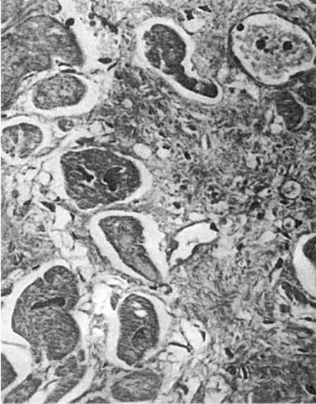
Connective tissue regeneration begins with the proliferation of young mesenchymal elements and new formation of microvessels. Young connective tissue, rich in cells and thin-walled vessels, is formed, which has a characteristic appearance. This is a juicy dark red fabric with a granular surface, as if strewn with large granules, which was the basis for calling it granulation tissue. Granules are loops of newly formed thin-walled vessels protruding above the surface, which form the basis of granulation tissue. Between the vessels there are many undifferentiated lymphocyte-like connective tissue cells, leukocytes, plasma cells and mast cells (Fig. 83). What happens next is maturation granulation tissue, which is based on the differentiation of cellular elements, fibrous structures, and blood vessels. The number of hematogenous elements decreases, and fibroblasts increase. In connection with the synthesis of collagen by fibroblasts, argyrophilic(see Fig. 83), and then collagen fibers. The synthesis of glycosaminoglycans by fibroblasts serves to form
main substance connective tissue. As fibroblasts mature, the number of collagen fibers increases and they are grouped into bundles; At the same time, the number of vessels decreases, they differentiate into arteries and veins. The maturation of granulation tissue ends with the formation coarse fibrous scar tissue.
New formation of connective tissue occurs not only when it is damaged, but also when other tissues are incompletely regenerated, as well as during organization (encapsulation), wound healing, and productive inflammation.
The maturation of granulation tissue may have one or another deviations. Inflammation developing in granulation tissue leads to a delay in its maturation,
 Rice. 83. Granulation tissue. Between the thin-walled vessels there are many undifferentiated connective tissue cells and argyrophilic fibers. Silver impregnation
and excessive synthetic activity of fibroblasts leads to excessive formation of collagen fibers, followed by pronounced hyalinosis. In such cases, scar tissue appears in the form of a tumor-like formation of a bluish-red color, which rises above the surface of the skin in the form keloid. Keloid scars form after various traumatic skin lesions, especially after burns.
Regeneration of adipose tissue occurs due to the new formation of connective tissue cells, which turn into fat cells (adipocytes) through the accumulation of lipids in the cytoplasm. Fat cells are folded into lobules, between which there are connective tissue layers with vessels and nerves. Regeneration of adipose tissue can also occur from nucleated remnants of the cytoplasm of fat cells.
Bone tissue regeneration in case of bone fracture, it largely depends on the degree of bone destruction, correct reposition of bone fragments, local conditions (circulatory conditions, inflammation, etc.). At uncomplicated bone fracture, when bone fragments are immobile, may occur primary bone union(Fig. 84). It begins with the ingrowth of young mesenchymal elements and vessels into the area of the defect and hematoma between bone fragments. There is a so-called preliminary connective tissue callus, in which bone formation immediately begins. It is associated with activation and proliferation osteoblasts in the damaged area, but primarily in the periostat and endostat. Slightly calcified bone beams appear in the osteogenic fibroreticular tissue, the number of which increases.
Formed preliminary callus. Subsequently, it matures and turns into mature lamellar bone - this is how
 Rice. 84. Primary bone fusion. Intermediary callus (shown by an arrow), fusing bone fragments (according to G.I. Lavrishcheva)
final callus, which in its structure differs from bone tissue only in the random arrangement of bone crossbars. After the bone begins to perform its function and a static load appears, the newly formed tissue undergoes restructuring with the help of osteoclasts and osteoblasts, bone marrow appears, and vascularization and innervation are restored. If local conditions for bone regeneration are violated (circulatory disorders), mobility of fragments, extensive diaphyseal fractures occur secondary bone fusion(Fig. 85). This type of bone fusion is characterized by the formation between bone fragments first cartilage tissue, on the basis of which bone tissue is built. Therefore, with secondary bone fusion they speak of preliminary osteochondral callus, which eventually develops into mature bone. Secondary bone fusion, compared to primary fusion, is much more common and takes longer.
At unfavorable conditions bone regeneration may be impaired. Thus, when a wound becomes infected, bone regeneration is delayed. Bone fragments, which during the normal course of the regenerative process serve as a frame for newly formed bone tissue, under conditions of suppuration of the wound, support inflammation, which inhibits regeneration. Sometimes the primary osteochondral callus does not differentiate into a bone callus. In these cases, the ends of the broken bone remain mobile, and a false joint. Excessive production of bone tissue during regeneration leads to the appearance of bone spurs - exostoses.
Regeneration of cartilage tissue unlike bone, it usually occurs incompletely. Only small defects can be replaced by newly formed tissue due to the cambial elements of the perichondrium - chondroblasts. These cells create the ground substance of cartilage, then develop into mature cartilage cells. Large cartilage defects are replaced by scar tissue.
Regeneration of muscle tissue, its capabilities and forms vary depending on the type of fabric. Smooth Muscles, whose cells have the ability to undergo mitosis and amitosis, can regenerate quite completely with minor defects. Significant areas of smooth muscle damage are replaced by scar, while the remaining muscle fibers undergo hypertrophy. New formation of smooth muscle fibers can occur through transformation (metaplasia) of connective tissue elements. This is how bundles of smooth muscle fibers are formed in pleural adhesions, in thrombi undergoing organization, and in vessels during their differentiation.
Striated muscles regenerate only if the sarcolemma is preserved. Inside the tubes from the sarcolemma, regeneration of its organelles occurs, resulting in the appearance of cells called myoblasts. They elongate, the number of nuclei in them increases, in the sarcoplasm
 Rice. 85. Secondary bone fusion (according to G.I. Lavrishcheva):
a - osteochondral periosteal callus; a section of bone tissue among cartilaginous tissue (microscopic picture); b - periosteal osteochondral callus (histotopogram 2 months after surgery): 1 - bone part; 2 - cartilaginous part; 3 - bone fragments; c - periosteal callus, fusing displaced bone fragments
myofibrils differentiate, and the sarcolemmal tubes transform into striated muscle fibers. Regeneration of skeletal muscles may also be associated with satellite cells, which are located under the sarcolemma, i.e. inside the muscle fiber, and are cambial. In case of injury, satellite cells begin to rapidly divide, then undergo differentiation and ensure the restoration of muscle fibers. If, when a muscle is damaged, the integrity of the fibers is disrupted, then flask-shaped protrusions appear at the ends of their breaks, which contain a large number of nuclei and are called muscle kidneys. In this case, restoration of fiber continuity does not occur. The rupture site is filled with granulation tissue, which turns into a scar (muscle callus). Regeneration heart muscles if it is damaged, as with damage to the striated muscles, it ends with scarring of the defect. However, in the remaining muscle fibers, intense hyperplasia of ultrastructures occurs, which leads to hypertrophy of the fibers and restoration of organ function (see Fig. 81).
Epithelial regeneration is carried out in most cases quite completely, since it has a high regenerative ability. Regenerates especially well cover epithelium. Recovery stratified squamous keratinizing epithelium possible even with fairly large skin defects. During the regeneration of the epidermis at the edges of the defect, increased proliferation of cells of the germinal (cambial) and germinal (Malpighian) layer occurs. The resulting epithelial cells first cover the defect in one layer. Subsequently, the layer of epithelium becomes multilayered, its cells differentiate, and it acquires all the signs of the epidermis, including the germinal, granular, shiny (on the soles and palmar surface of the hands) and stratum corneum. When the regeneration of the skin epithelium is impaired, non-healing ulcers are formed, often with the growth of atypical epithelium at their edges, which can serve as the basis for the development of skin cancer.
Covering epithelium of mucous membranes (multilayered squamous non-keratinized, transitional, single-layered prismatic and multinucleated ciliated) regenerates in the same way as multi-layered squamous keratinized. The mucosal defect is restored due to the proliferation of cells lining the crypts and excretory ducts of the glands. Undifferentiated flattened epithelial cells first cover the defect with a thin layer (Fig. 86), then the cells take on the shape characteristic of the cellular structures of the corresponding epithelial lining. In parallel, the glands of the mucous membrane (for example, tubular glands of the intestine, endometrial glands) are partially or completely restored.
Mesothelium regeneration peritoneum, pleura and pericardial sac is carried out by dividing the surviving cells. Relatively large cubic cells appear on the surface of the defect, which then flatten. For small defects, the mesothelial lining is restored quickly and completely.
The condition of the underlying connective tissue is important for the restoration of the integumentary epithelium and mesothelium, since epithelization of any defect is possible only after filling it with granulation tissue.
Regeneration of specialized organ epithelium(liver, pancreas, kidneys, endocrine glands, pulmonary alveoli) is carried out according to the type regenerative hypertrophy: in areas of damage, the tissue is replaced by a scar, and along its periphery hyperplasia and hypertrophy of parenchyma cells occur. IN liver the area of necrosis is always subject to scarring, but in the rest of the organ there is intense new cell formation, as well as hyperplasia of intracellular structures, which is accompanied by their hypertrophy. As a result, the original mass and function of the organ are quickly restored. The regenerative capabilities of the liver are almost limitless. In the pancreas, regenerative processes are well expressed both in the exocrine sections and in the pancreatic islets, and the epithelium of the exocrine glands becomes the source of restoration of the islets. IN kidneys with necrosis of the tubular epithelium, the surviving nephrocytes multiply and the tubules are restored, but only when the tubular basement membrane is preserved. When it is destroyed (tubulorrhexis), the epithelium is not restored and the tubule is replaced by connective tissue. The dead tubular epithelium is not restored even in the case when the vascular glomerulus dies simultaneously with the tubule. In this case, scar connective tissue grows in the place of the dead nephron, and the surrounding nephrons undergo regenerative hypertrophy. In the glands internal secretion restoration processes are also represented by incomplete regeneration. IN lung after removal of individual lobes, hypertrophy and hyperplasia of tissue elements occurs in the remaining part. Regeneration of specialized epithelium of organs can proceed atypically, which leads to the proliferation of connective tissue, structural restructuring and deformation of organs; in such cases we talk about cirrhosis (liver cirrhosis, nephrocirrhosis, pneumocirrhosis).
Regeneration of different parts of the nervous system happens ambiguously. IN head And spinal cord neoplasms of ganglion cells do not pro-
 Rice. 86. Regeneration of epithelium in the bottom of a chronic gastric ulcer
occurs and when they are destroyed, restoration of function is possible only through intracellular regeneration of surviving cells. Neuroglia, especially microglia, are characterized by a cellular form of regeneration, therefore defects in the tissue of the brain and spinal cord are usually filled with proliferating neuroglial cells - so-called glial (gliotic) scarring. If damaged vegetative nodes Along with hyperplasia of cell ultrastructures, their new formation also occurs. In case of violation of integrity peripheral nerve regeneration occurs due to the central segment, which has retained its connection with the cell, while the peripheral segment dies. The multiplying cells of the Schwann sheath of the dead peripheral segment of the nerve are located along it and form a sheath - the so-called Büngner cord, into which regenerating axial cylinders from the proximal segment grow. Regeneration of nerve fibers ends with their myelination and restoration of nerve endings. Regenerative hyperplasia receptors, pericellular synaptic devices and effectors are sometimes accompanied by hypertrophy of their terminal apparatus. If nerve regeneration is disrupted for one reason or another (significant divergence of parts of the nerve, development of an inflammatory process), then at the site of its interruption a scar is formed in which the regenerated axial cylinders of the proximal segment of the nerve are randomly located. Similar growths occur at the ends of cut nerves in the stump of a limb after amputation. Such growths formed by nerve fibers and fibrous tissue are called amputation neuromas.
Wound healing
Wound healing proceeds according to the laws of reparative regeneration. The rate of wound healing and its outcomes depend on the degree and depth of wound damage, the structural features of the organ, the general condition of the body, and the treatment methods used. According to I.V. Davydovsky, the following types of wound healing are distinguished: 1) direct closure of the epithelial defect; 2) healing under the scab; 3) wound healing by primary intention; 4) wound healing by secondary intention, or wound healing through suppuration.
Direct closure of epithelial defect- this is the simplest healing, which consists of the creeping of the epithelium over the surface defect and covering it with an epithelial layer. Observed on the cornea, mucous membranes healing under the scab concerns small defects, on the surface of which a drying crust (scab) of clotted blood and lymph quickly appears; the epidermis is restored under a crust, which disappears 3-5 days after injury.
Healing by primary intention (per rimam intentionem) observed in wounds with damage not only to the skin, but also to the underlying tissue,
and the edges of the wound are smooth. The wound is filled with clots of spilled blood, which protects the edges of the wound from dehydration and infection. Under the influence of proteolytic enzymes of neutrophils, partial lysis of blood clotting and tissue detritus occurs. Neutrophils die and are replaced by macrophages, which phagocytize red blood cells and the remains of damaged tissue; Hemosiderin is found at the edges of the wound. Part of the contents of the wound is removed on the first day of the wound along with exudate independently or during wound treatment - primary cleansing. On the 2-3rd day, fibroblasts and newly formed capillaries appear at the edges of the wound, growing towards each other, granulation tissue, the layer of which does not reach large sizes during primary tension. By the 10-15th day it fully matures, the wound defect is epithelialized and the wound heals with a delicate scar. In a surgical wound, healing by primary intention is accelerated due to the fact that its edges are tightened with threads of silk or catgut, around which giant cells of foreign bodies accumulate that dissolve them and do not interfere with healing.
Healing by secondary intention (per secundam intentionem), or healing through suppuration (or healing through granulation - per granulationem), It is usually observed with extensive wounds, accompanied by crushing and necrosis of tissue, penetration of foreign bodies and microbes into the wound. Hemorrhages and traumatic swelling of the wound edges occur at the wound site, and signs of demarcation quickly appear. purulent inflammation at the border with dead tissue, melting of necrotic masses. During the first 5-6 days, necrotic masses are rejected - secondary cleansing of the wound, and granulation tissue begins to develop at the edges of the wound. granulation tissue, filling the wound, consists of 6 layers passing into each other (Anichkov N.N., 1951): superficial leukocyte-necrotic layer; superficial layer of vascular loops, layer of vertical vessels, maturing layer, layer of horizontally located fibroblasts, fibrous layer. The maturation of granulation tissue during wound healing by secondary intention is accompanied by epithelial regeneration. However, with this type of wound healing, a scar always forms at its site.
REGENERATION
restoration by the body of lost parts at one stage or another life cycle. Regeneration usually occurs in the event of damage or loss of an organ or part of the body. However, in addition to this, restoration and renewal processes constantly occur in every organism throughout its life. In humans, for example, the outer layer of skin is constantly renewed. Birds periodically shed their feathers and grow new ones, and mammals change their fur. Deciduous trees lose leaves every year and are replaced with fresh ones. Such regeneration, usually not associated with damage or loss, is called physiological. Regeneration that occurs after damage or loss of any part of the body is called reparative. Here we will consider only reparative regeneration. Reparative regeneration can be typical or atypical. In typical regeneration, the lost part is replaced by the development of exactly the same part. The cause of the loss may be an external force (for example, amputation), or the animal may deliberately tear off part of its body (autotomy), like a lizard breaking off part of its tail to escape an enemy. With atypical regeneration, the lost part is replaced by a structure that differs from the original quantitatively or qualitatively. The regenerated limb of a tadpole may have fewer toes than the original one, and a shrimp may grow an antenna instead of an amputated eye.
REGENERATION IN ANIMALS
The ability to regenerate is widespread among animals. Generally speaking, lower animals are more often capable of regeneration than more complex, highly organized forms. Thus, among invertebrates there are many more species capable of restoring lost organs than among vertebrates, but only in some of them is it possible to regenerate an entire individual from a small fragment. Nevertheless general rule the decrease in the ability to regenerate with increasing complexity of the organism cannot be considered absolute. Such primitive animals as ctenophores and rotifers are practically incapable of regeneration, but in much more complex crustaceans and amphibians this ability is well expressed; Other exceptions are known. Some closely related animals differ greatly in this respect. Yes, y a new individual can completely regenerate from a small piece of the body, while leeches are unable to restore one lost organ. In tailed amphibians, a new limb is formed in place of the amputated limb, but in the frog, the stump simply heals and no new growth occurs. Many invertebrates are capable of regenerating large parts of their body. In sponges, hydroid polyps, flat, ribbon and annelids, bryozoans, echinoderms and tunicates, a whole organism can be regenerated from a small fragment of the body. Particularly noteworthy is the ability to regenerate in sponges. If the body of an adult sponge is pressed through the mesh fabric, then all the cells will separate from each other, as if sifted through a sieve. If you then place all these individual cells in water and carefully, thoroughly mix, completely destroying all the connections between them, then after some time they begin to gradually come closer together and reunite, forming a whole sponge, similar to the previous one. This involves a kind of "recognition" at the cellular level, as evidenced by the following experiment. Sponges of three different species were separated into separate cells in the manner described and mixed thoroughly. At the same time, it was discovered that the cells of each species are able to “recognize” the cells of their own species in the total mass and reunite only with them, so that as a result, not one, but three new sponges were formed, similar to the three original ones.
The tapeworm, which is many times longer than it is wide, can recreate an entire individual from any part of its body. It is theoretically possible, by cutting one worm into 200,000 pieces, to obtain 200,000 new worms from it as a result of regeneration. From one ray of a starfish, an entire star can regenerate.

Mollusks, arthropods and vertebrates are not able to regenerate a whole individual from one fragment, however, in many of them the lost organ is restored. Some resort to autotomy if necessary. Birds and mammals, as the most evolutionarily advanced animals, are less capable of regeneration than others. In birds, it is possible to replace feathers and some parts of the beak. Mammals can restore their integument, claws, and partly their liver; they are also capable of healing wounds, and deer are capable of growing new antlers to replace those shed.
Regeneration processes. Two processes are involved in regeneration in animals: epimorphosis and morphallaxis. In epimorphic regeneration, the lost part of the body is restored due to the activity of undifferentiated cells. These embryonic-like cells accumulate under the wounded epidermis at the cut surface, where they form the primordium, or blastema. Blastema cells gradually multiply and transform into the tissue of a new organ or body part. In morphallaxis, other tissues of the body or organ are directly transformed into the structures of the missing part. In hydroid polyps, regeneration occurs mainly through morphallaxis, while in planarians, both epimorphosis and morphallaxis are simultaneously involved in it. Regeneration by blastema formation is widespread in invertebrates and plays a particularly important role in organ regeneration in amphibians. There are two theories of the origin of blastema cells: 1) blastema cells originate from “reserve cells”, i.e. cells that remained unused during embryonic development and were distributed among different organs of the body; 2) tissues, the integrity of which was violated during amputation, “dedifferentiate” in the area of the incision, i.e. disintegrate and transform into individual blastema cells. Thus, according to the “reserve cell” theory, the blastema is formed from cells that remained embryonic, which migrate from different parts of the body and accumulate at the cut surface, and according to the “dedifferentiated tissue” theory, blastema cells originate from cells of damaged tissues. There is enough data to support both one and the other theory. For example, in planarians, reserve cells are more sensitive to x-rays than cells of differentiated tissue; therefore, they can be destroyed by strictly dosing radiation so as not to damage normal planarian tissue. Individuals irradiated in this way survive, but lose their ability to regenerate. However, if only the anterior half of the planarian body is irradiated and then cut, then regeneration occurs, although with some delay. The delay indicates that the blastema is formed from reserve cells migrating to the cut surface from the non-irradiated half of the body. The migration of these reserve cells throughout the irradiated part of the body can be observed under a microscope. Similar experiments showed that in the newt, limb regeneration occurs due to blastema cells of local origin, i.e. due to dedifferentiation of damaged stump tissues. If, for example, you irradiate the entire newt larva except, say, the right forelimb, and then amputate that limb at the level of the forearm, the animal will grow a new forelimb. It is obvious that the blastema cells necessary for this come precisely from the stump of the forelimb, since the rest of the body has been irradiated. Moreover, regeneration occurs even if the entire larva is irradiated, with the exception of a 1 mm wide area on the right front leg, and then the latter is amputated by making an incision through this non-irradiated area. In this case, it is quite clear that the blastema cells come from the cut surface, since the entire body, including the right foreleg, was deprived of the ability to regenerate. The described processes were analyzed using modern methods. An electron microscope allows you to observe changes in damaged and regenerating tissues in all details. Dyes have been created that reveal certain chemicals contained in cells and tissues. Histochemical methods (using dyes) make it possible to judge the biochemical processes occurring during the regeneration of organs and tissues.
Polarity. One of the most mysterious problems in biology is the origin of polarity in organisms. From the spherical egg of a frog, a tadpole develops, which from the very beginning has a head with a brain, eyes and mouth at one end of the body, and a tail at the other. Similarly, if you cut the body of a planarian into individual fragments, a head develops at one end of each fragment and a tail at the other. In this case, the head is always formed at the anterior end of the fragment. Experiments clearly show that the planarian has a gradient of metabolic (biochemical) activity along the anterior-posterior axis of its body; in this case, the highest activity is at the very anterior end of the body, and towards the posterior end the activity gradually decreases. In any animal, the head is always formed at the end of the fragment where metabolic activity is higher. If the direction of the metabolic activity gradient in an isolated planarian fragment is reversed, then the formation of the head will occur at the opposite end of the fragment. The gradient of metabolic activity in the body of planarians reflects the existence of some more important physicochemical gradient, the nature of which is still unknown. In the regenerating limb of a newt, the polarity of the newly formed structure appears to be determined by the preserved stump. For reasons that still remain unclear, only structures located distal to the wound surface are formed in the regenerating organ, and those located more proximally (closer to the body) never regenerate. So, if the hand of a newt is amputated, and the remaining part of the forelimb is inserted with the cut end into the body wall and this distal (distant from the body) end is allowed to take root in a new, unusual place for it, then the subsequent cutting of this upper limb near the shoulder (freeing it from the connection with the shoulder) leads to the regeneration of the limb with a full set of distal structures. At the time of cutting, such a limb has the following parts (starting from the wrist, fused with the body wall): wrist, forearm, elbow and distal half of the shoulder; then, as a result of regeneration, the following appear: another distal half of the shoulder, elbow, forearm, wrist and hand. Thus, the inverted (upside down) limb regenerated all parts located distal to the wound surface. This striking phenomenon indicates that the tissues of the stump (in this case the limb stump) control the regeneration of the organ. The task of further research is to find out exactly what factors control this process, what stimulates regeneration and what causes the cells that ensure regeneration to accumulate on the wound surface. Some scientists believe that damaged tissue releases some kind of chemical "wound factor." However, highlight Chemical substance, specific for wounds, has not yet been successful.
REGENERATION IN PLANTS
The widespread occurrence of regeneration in the plant kingdom is due to the preservation of meristems (tissues consisting of dividing cells) and undifferentiated tissues. In most cases, regeneration in plants is, in essence, one of the forms of vegetative propagation. Thus, at the tip of a normal stem there is an apical bud, which ensures the continuous formation of new leaves and the growth of the stem in length throughout the life of the plant. If this bud is cut off and kept moist, new roots often develop from the parenchyma cells present in it or from the callus formed on the surface of the cut; the bud continues to grow and gives rise to a new plant. The same thing happens in nature when a branch breaks off. The lashes and stolons are separated as a result of the death of old sections (internodes). In the same way, the rhizomes of iris, wolf's foot or ferns are divided, forming new plants. Typically, tubers, such as potato tubers, continue to live after the underground stem on which they grew has died; with the onset of a new growing season, they can give rise to their own roots and shoots. In bulbous plants, such as hyacinths or tulips, shoots form at the base of the bulb scales and can in turn form new bulbs, which eventually produce roots and flowering stems, i.e. become independent plants. In some lilies, aerial bulbs form in the axils of the leaves, and in a number of ferns, brood buds grow on the leaves; at some point they fall to the ground and resume growth. Roots are less capable of forming new parts than stems. For this, the dahlia tuber needs a bud that forms at the base of the stem; however, sweet potatoes can give rise to a new plant from a bud formed by a root cone. Leaves are also capable of regeneration. In some species of ferns, for example, in the fern (Camptosorus), the leaves are very elongated and have the appearance of long hair-like formations ending in a meristem. From this meristem the embryo develops with rudimentary stem, roots and leaves; if the tip of the parent plant's leaf bends down and touches the soil or moss, the bud begins to grow. The new plant separates from the parent after the depletion of this hair-like formation. The leaves of the succulent houseplant Kalanchoe bear well-developed plantlets at the edges that fall off easily. New shoots and roots form on the surface of begonia leaves. Special bodies called embryonic buds develop on the leaves of some club mosses (Lycopodium) and liverworts (Marchantia); falling to the ground, they take root and form new mature plants. Many algae reproduce successfully by breaking into fragments under the impact of waves.
see also PLANT SYSTEMATICS. LITERATURE Mattson P. Regeneration - present and future. M., 1982 Gilbert S. Developmental biology, vol. 1-3. M., 1993-1995
Collier's Encyclopedia. - Open Society. 2000 .
Synonyms:See what "REGENERATION" is in other dictionaries:
REGENERATION- REGENERATION, the process of formation of a new organ or tissue in place of a part of the body that was removed in one way or another. Very often R. is defined as the process of restoring what has been lost, that is, the formation of an organ similar to the removed one. This... ... Great Medical Encyclopedia
- (late lat., from lat. re again, again, and genus, eris genus, generation). Revival, renewal, restoration of what was destroyed. In a figurative meaning: a change for the better. Dictionary of foreign words included in the Russian language.... ... Dictionary of foreign words of the Russian language
REGENERATION, in biology, the body’s ability to replace one of the lost parts. The term regeneration also refers to a form of Asexual Reproduction in which a new individual arises from a separated part of the mother's body... Scientific and technical encyclopedic dictionary
Restoration, recovery; compensation, regeneration, renewal, heteromorphosis, pettencoferation, revival, morphallaxis Dictionary of Russian synonyms. regeneration noun, number of synonyms: 11 compensation (20) ... Synonym dictionary
1) restoration, using certain physicochemical processes, of the original composition and properties of waste products for their reuse. In military affairs, air regeneration has become widespread (especially on underwater... ... Marine Dictionary
Regeneration- – returning the used product to its original properties. [Terminological dictionary of concrete and reinforced concrete. FSUE "Research Center "Construction" NIIZHB named after. A. A. Gvozdeva, Moscow, 2007, 110 pp.] Regeneration - restoration of waste... ... Encyclopedia of terms, definitions and explanations of building materials
REGENERATION- (1) restoration of the original properties and composition of waste materials (water, air, oils, rubber, etc.) for their reuse. It is carried out with the help of certain physical chem. processes in special regenerator devices. Wide... ... Big Polytechnic Encyclopedia
Academy of Regenerative Medicine is a training, research, medical, recreational, regenerative and gerontology (rejuvenation) institution, founded in Swiebodzice, Poland in 2010. In a short period of time, the Academy of Regenerative Medicine gained worldwide fame and became one of the leading medical centers in Europe. Patients from 30 countries (the USA, Canada, Australia, Israel, EU, CIS, Asian and African countries) have already undergone successfully their treatment and regeneration in our center. The Academy of Regenerative Medicine is not only a leader in preventing and treating many chronic incurable and genetic diseases but the only center in the world in which the integrated method of body regeneration with the help of simple harmless natural techniques is implemented and widely used. A promising new direction in complementary and regenerative medicine was developed, proven and implemented in practice in our center.
The invaluable advantage of this technique is the rapid restoration, regeneration and rejuvenation of the body in so much that our patients in any condition and at any age almost completely stop taking any medications, including painkillers, food supplements. They also stop restricting their food and keeping to any diet. THEY LIVE A FULL LIFE WITHOUT DISEASES AND MEDICATIONS!
The Academy of Regenerative Medicine works on the basis of the author’s method of Aliaksandr Haretski “The method of human organ regeneration, biological body rejuvenation, integrated healing of chronic “incurable” diseases and aging with the help of regenerative medicine techniques.”
You can get acquainted with the basic elements of our method on the official website of our Academy www.acadregmed.com We have posted lots of practical information there. You can learn how to effectively cleanse the body of harmful toxins and parasites, how to eliminate the main causes of diseases and boost the immune system. You can also learn how to launch the mechanisms of body regeneration and self-healing in sick people with various severe diseases.


OUR REGENERATION METHOD IS UNPARALLELED ANYWHERE ELSE IN THE WORLD!
Our Academy of Regenerative Medicine is the only health care institution in the world in which only harmless natural methods of treatment and our own know-how developments are used. We are in Poland and Russia.
UNIQUE POSITIVE SIDE EFFECTS!
The uniqueness of our method is that it allows us to stop the aging process and replace the name of many “incurable diseases” with “curable diseases”. The use of this technique offers an opportunity to give people a new lease on life at minimal cost. It's universal and harmless. It has virtually no contraindications. It can be effectively used not only for prevention and treatment of many diseases but also for body rejuvenation at any age. The positive side effect, which manifests itself as the regeneration and rejuvenation not only of the skin but also of the whole human body, means that this is the best place on earth to relax and improve your health and beauty. After undergoing treatment in our Academy, every person looks and feels healthy and many years younger without any plastic surgery. We invite you to see it for yourself!
PROFESSIONALISM AND QUALITY!
The Academy of Regenerative Medicine has earned high accolades and recognition from its patients all over the world owing to our highly professional specialists, our high level of customer service and the possibility of comprehensive disease treatment. The staff of our center speaks Polish, Russian, English and many other languages fluently and provides high-quality assistance to patients from any country.
REASONABLE PRICES!
The cost of our services, compared with the cost of services of other medical centers in European countries, the USA, Japan and even China, is tens or even hundreds of times lower than when using other methods of treatment of incurable diseases. Our prices are lower but the effectiveness is higher!
A HIGH QUALITY OF LIFE AFTER THE BODY REGENERATION!
The important advantage of our work is a high quality of life of our patients after their treatment in our center with healthy organs and rejuvenated cells in the whole body, with a strong immune system, without diseases and drugs.
THE SALUBRIOUS CLIMATE!
Our Academy is located in the foothills area in the south-western part of Poland. The main advantages of this region are clean air and a mild climate with favorable weather conditions all the year round.
COMFORTABLE ACCOMMODATION!
We offer comfortable self-catering accommodation with home atmosphere in well-equipped one bedroom and two bedroom apartments with a bathroom and a kitchen in our center. It allows our patients not to feel like staying in hospital and provides psychological comfort.
HOW TO CHOOSE A COURSE OF REGENERATIVE THERAPY CORRECTLY?
After getting acquainted with the information posted on our website many people think that we are wizards and within a week we can eliminate big problems that have existed for many years. Sometimes very quick fantastic results are achieved in our center - God works miracles, but this is an exception. In most cases, we and our patient must work hard and long hours with the help of God in order to achieve a goal. Our task is to cleanse the body and make it replace its old, sick and damaged cells with young and healthy ones. The regeneration process of the very damaged and weakened body is very time consuming. We can only recommend the right course of regenerative therapy for you. The choice always remains with the patient and depends on his faith, desires and possibilities! And thereby the result depends on your choice!
YOUR DESTINY LIES IN YOUR HANDS!


SO AGAIN, HOW TO CHOOSE A COURSE OF REGENERATIVE THERAPY CORRECTLY?
After getting acquainted with the information posted on our website many people think that we are wizards and within a week we can eliminate big problems that have existed for many years. Sometimes very quick fantastic results are achieved in our center - God works miracles, but this is an exception. In most cases, we and our patient must work hard and long hours with the help of God in order to achieve a goal. Our task is to cleanse the body and make it replace its old, sick and damaged cells with young and healthy ones. The regeneration process of the very damaged and weakened body is very time consuming. We can only recommend the right course of regenerative therapy for you. The choice always remains with the patient and depends on his faith, desires and possibilities! And thereby the result depends on your choice!
REMEMBER! YOUR DESTINY LIES IN YOUR HANDS!


The content of the article
REGENERATION, restoration by the body of lost parts at one or another stage of the life cycle. Regeneration usually occurs in the event of damage or loss of an organ or part of the body. However, in addition to this, restoration and renewal processes constantly occur in every organism throughout its life. In humans, for example, the outer layer of skin is constantly renewed. Birds periodically shed their feathers and grow new ones, and mammals change their fur. Deciduous trees lose leaves every year and are replaced with fresh ones. Such regeneration, usually not associated with damage or loss, is called physiological. Regeneration that occurs after damage or loss of any part of the body is called reparative. Here we will consider only reparative regeneration.
Reparative regeneration can be typical or atypical. In typical regeneration, the lost part is replaced by the development of exactly the same part. The cause of the loss may be an external force (for example, amputation), or the animal may deliberately tear off part of its body (autotomy), like a lizard breaking off part of its tail to escape an enemy. With atypical regeneration, the lost part is replaced by a structure that differs from the original quantitatively or qualitatively. The regenerated limb of a tadpole may have fewer toes than the original one, and a shrimp may grow an antenna instead of an amputated eye.
REGENERATION IN ANIMALS
The ability to regenerate is widespread among animals. Generally speaking, lower animals are more often capable of regeneration than more complex, highly organized forms. Thus, among invertebrates there are many more species capable of restoring lost organs than among vertebrates, but only in some of them is it possible to regenerate an entire individual from a small fragment. Nevertheless, the general rule that the ability to regenerate decreases with increasing complexity of the organism cannot be considered absolute. Such primitive animals as ctenophores and rotifers are practically incapable of regeneration, but in much more complex crustaceans and amphibians this ability is well expressed; Other exceptions are known. Some closely related animals differ greatly in this respect. Thus, in an earthworm, a new individual can completely regenerate from a small piece of its body, while leeches are unable to restore one lost organ. In tailed amphibians, a new limb is formed in place of the amputated limb, but in the frog, the stump simply heals and no new growth occurs.
Many invertebrates are capable of regenerating large parts of their body. In sponges, hydroid polyps, flatworms, tapeworms and annelids, bryozoans, echinoderms and tunicates, a whole organism can regenerate from a small fragment of the body. Particularly noteworthy is the ability to regenerate in sponges. If the body of an adult sponge is pressed through the mesh fabric, then all the cells will separate from each other, as if sifted through a sieve. If you then place all these individual cells in water and carefully, thoroughly mix, completely destroying all the connections between them, then after some time they begin to gradually come closer together and reunite, forming a whole sponge, similar to the previous one. This involves a kind of “recognition” at the cellular level, as evidenced by the following experiment. Sponges of three different species were separated into separate cells in the manner described and mixed thoroughly. At the same time, it was discovered that the cells of each species are able to “recognize” the cells of their own species in the total mass and reunite only with them, so that as a result, not one, but three new sponges were formed, similar to the three original ones.
The tapeworm, which is many times longer than it is wide, can recreate an entire individual from any part of its body. It is theoretically possible, by cutting one worm into 200,000 pieces, to obtain 200,000 new worms from it as a result of regeneration. From one ray of a starfish, an entire star can regenerate.
Mollusks, arthropods and vertebrates are not able to regenerate a whole individual from one fragment, however, in many of them the lost organ is restored. Some resort to autotomy if necessary. Birds and mammals, as the most evolutionarily advanced animals, are less capable of regeneration than others. In birds, it is possible to replace feathers and some parts of the beak. Mammals can restore their integument, claws, and partly their liver; they are also capable of healing wounds, and deer are capable of growing new antlers to replace those shed.
Regeneration processes.
Two processes are involved in regeneration in animals: epimorphosis and morphallaxis. In epimorphic regeneration, the lost part of the body is restored due to the activity of undifferentiated cells. These embryonic-like cells accumulate under the wounded epidermis at the cut surface, where they form the primordium, or blastema. Blastema cells gradually multiply and transform into the tissue of a new organ or body part. In morphallaxis, other tissues of the body or organ are directly transformed into the structures of the missing part. In hydroid polyps, regeneration occurs mainly through morphallaxis, while in planarians, both epimorphosis and morphallaxis are simultaneously involved in it.
Regeneration by blastema formation is widespread in invertebrates and plays a particularly important role in organ regeneration in amphibians. There are two theories of the origin of blastema cells: 1) blastema cells originate from “reserve cells”, i.e. cells that remained unused during embryonic development and were distributed among different organs of the body; 2) tissues, the integrity of which was violated during amputation, “dedifferentiate” in the area of the incision, i.e. disintegrate and transform into individual blastema cells. Thus, according to the “reserve cell” theory, the blastema is formed from cells that remained embryonic, which migrate from different parts of the body and accumulate near the cut surface, and according to the “dedifferentiated tissue” theory, blastema cells originate from cells of damaged tissues.
There is enough data to support both one and the other theory. For example, in planarians, reserve cells are more sensitive to X-rays than cells of differentiated tissue; therefore, they can be destroyed by strictly dosing radiation so as not to damage normal planarian tissue. Individuals irradiated in this way survive, but lose their ability to regenerate. However, if only the anterior half of the planarian body is irradiated and then cut, then regeneration occurs, although with some delay. The delay indicates that the blastema is formed from reserve cells migrating to the cut surface from the non-irradiated half of the body. The migration of these reserve cells throughout the irradiated part of the body can be observed under a microscope.
Similar experiments showed that in the newt, limb regeneration occurs due to blastema cells of local origin, i.e. due to dedifferentiation of damaged stump tissues. If, for example, you irradiate the entire newt larva except, say, the right forelimb, and then amputate that limb at the level of the forearm, the animal will grow a new forelimb. It is obvious that the blastema cells necessary for this come precisely from the stump of the forelimb, since the rest of the body has been irradiated. Moreover, regeneration occurs even if the entire larva is irradiated, with the exception of a 1 mm wide area on the right front leg, and then the latter is amputated by making an incision through this non-irradiated area. In this case, it is quite clear that the blastema cells come from the cut surface, since the entire body, including the right foreleg, was deprived of the ability to regenerate.
The described processes were analyzed using modern methods. An electron microscope allows you to observe changes in damaged and regenerating tissues in all details. Dyes have been created that reveal certain chemicals contained in cells and tissues. Histochemical methods (using dyes) make it possible to judge the biochemical processes occurring during the regeneration of organs and tissues.
Polarity.
One of the most mysterious problems in biology is the origin of polarity in organisms. From the spherical egg of a frog, a tadpole develops, which from the very beginning has a head with a brain, eyes and mouth at one end of the body, and a tail at the other. Similarly, if you cut the body of a planarian into individual fragments, a head develops at one end of each fragment and a tail at the other. In this case, the head is always formed at the anterior end of the fragment. Experiments clearly show that the planarian has a gradient of metabolic (biochemical) activity along the anterior-posterior axis of its body; in this case, the highest activity is at the very anterior end of the body, and towards the posterior end the activity gradually decreases. In any animal, the head is always formed at the end of the fragment where metabolic activity is higher. If the direction of the metabolic activity gradient in an isolated planarian fragment is reversed, then the formation of the head will occur at the opposite end of the fragment. The gradient of metabolic activity in the body of planarians reflects the existence of some more important physicochemical gradient, the nature of which is still unknown.
In the regenerating limb of a newt, the polarity of the newly formed structure appears to be determined by the preserved stump. For reasons that still remain unclear, only structures located distal to the wound surface are formed in the regenerating organ, and those located more proximally (closer to the body) never regenerate. So, if the hand of a newt is amputated, and the remaining part of the forelimb is inserted with the cut end into the body wall and this distal (distant from the body) end is allowed to take root in a new, unusual place for it, then the subsequent cutting of this upper limb near the shoulder (freeing it from the connection with the shoulder) leads to the regeneration of the limb with a full set of distal structures. At the time of cutting, such a limb has the following parts (starting from the wrist, fused with the body wall): wrist, forearm, elbow and distal half of the shoulder; then, as a result of regeneration, the following appear: another distal half of the shoulder, elbow, forearm, wrist and hand. Thus, the inverted (upside down) limb regenerated all parts located distal to the wound surface. This striking phenomenon indicates that the tissues of the stump (in this case the limb stump) control the regeneration of the organ. The task of further research is to find out exactly what factors control this process, what stimulates regeneration and what causes the cells that ensure regeneration to accumulate on the wound surface. Some scientists believe that damaged tissue releases some kind of chemical “wound factor.” However, it has not yet been possible to isolate a chemical substance specific to wounds.
REGENERATION IN PLANTS
The widespread occurrence of regeneration in the plant kingdom is due to the preservation of meristems (tissues consisting of dividing cells) and undifferentiated tissues. In most cases, regeneration in plants is, in essence, one of the forms of vegetative propagation. Thus, at the tip of a normal stem there is an apical bud, which ensures the continuous formation of new leaves and the growth of the stem in length throughout the life of the plant. If this bud is cut off and kept moist, new roots often develop from the parenchyma cells present in it or from the callus formed on the surface of the cut; the bud continues to grow and gives rise to a new plant. The same thing happens in nature when a branch breaks off. The lashes and stolons are separated as a result of the death of old sections (internodes). In the same way, the rhizomes of iris, wolf's foot or ferns are divided, forming new plants. Typically, tubers, such as potato tubers, continue to live after the underground stem on which they grew has died; with the onset of a new growing season, they can give rise to their own roots and shoots. In bulbous plants, such as hyacinths or tulips, shoots form at the base of the bulb scales and can in turn form new bulbs, which eventually produce roots and flowering stems, i.e. become independent plants. In some lilies, aerial bulbs form in the axils of the leaves, and in a number of ferns, brood buds grow on the leaves; at some point they fall to the ground and resume growth.
Roots are less capable of forming new parts than stems. For this, the dahlia tuber needs a bud that forms at the base of the stem; however, sweet potatoes can give rise to a new plant from a bud formed by a root cone.
Leaves are also capable of regeneration. In some species of ferns, for example, the crooked fern ( Camptosorus), the leaves are highly elongated and have the appearance of long hair-like formations ending in a meristem. From this meristem the embryo develops with rudimentary stem, roots and leaves; if the tip of the parent plant's leaf bends down and touches the soil or moss, the bud begins to grow. The new plant separates from the parent after the depletion of this hair-like formation. The leaves of the succulent houseplant Kalanchoe bear well-developed plantlets at the edges that fall off easily. New shoots and roots form on the surface of begonia leaves. Special bodies called embryonic buds develop on the leaves of some club mosses (Lycopodium) and liverworts (Marchantia); falling to the ground, they take root and form new mature plants.
The human body is unique. The cells that make up all its organs are capable of dividing a certain number of times, thereby replacing dead ones. Of course, we cannot compare with lizards, who can grow a new tail in a few days, but we cannot be considered as completely devoid of the ability to self-heal. Within certain limits, the body experiences cell regeneration blood, skin, pancreas and even nerves. Yes, yes, I didn’t make a reservation - nerve cells have the ability to recover!
I, like many of you, was convinced of the opposite. Until the mid-20th century, there was a theory that a person is born with a certain supply of nerve cells, and throughout his life, he gradually spends this supply. In 1962, American professor Altman refuted this theory, proving that new nerve cells in the human brain are restored due to stem cells located around the ventricles of the cerebral hemispheres. Today, this discovery is successfully used to treat diseases accompanied by the death of brain neurons. To the area of the brain that needs to be restored, using special equipment stem cells are introduced.
I think many people have heard about the wonderful properties of stem cells. I will try to explain clearly what kind of “treasure” this is in the health of the body. Stem cells are embryonic cells that are the precursors of all body tissues. Under the influence of various conditions, they are able to turn into nerves, muscles and skin. The main reserve of stem cells in the body is the red bone marrow, which contains the so-called building cells, whose name is stromal. They constantly circulate in the blood.
If a “breakdown” occurs in any organ, stromal cells rush to the scene of the disaster and, under the influence of special substances, transform into the necessary cells. Unfortunately, their amount in the blood is not very large, so they can only cope with small “accidents” in the body.
Many scientists see stem cells as a way to prolong life and preserve the health of the human body. Work in this direction is underway, but science does not yet know how stem cells can be brought to life and forced to recreate lost tissue. I hope that in the near future the veil of secrecy will be lifted.
Over the years, whether we like it or not, skin aging occurs. The fact is that only the top layer of skin, the epidermis, has the ability to quickly renew and divide. As a rule, it is completely updated every 4 weeks. Alas, after 45 years this period is extended to 3 months.
In the deeper layer of the skin - the dermis - not all cells have the ability to restore, but only elastin and collagen fibers. The main cells in the dermis are fibroblasts. They are the ones who synthesize elastin and collagen. It is important to know that a decrease in the synthesis of elastin begins at the age of 25, and collagen - at the age of 30. Of course, we women, like many men, try to resist this process. The regenerative capabilities of the skin largely depend on genetics, but proper nutrition, as well as good nutrition, play an important role.