How is the cervix located? Before childbirth: Basic medical terms of childbirth
This project was led by a 25-year-old woman. She has never given birth and has no history of STDs. Each photograph was taken at approximately 10:00 p.m., starting on the first day of the menstrual cycle. Throughout this project, she used condoms as a contraceptive method and also to prevent semen from being released during the photo shoot. She did not use tampons during her period.
This cycle is 33 days, which is the norm. The follicular phase of her cycle lasts until about 20–21 days. Favorable days for fertilization last several days from the 13th to the 21st day with ovulation on the 20th day. The luteal phase is 13 days (12–16 days is normal).
The above is for this cycle. As you can see, after ovulation on about the 20th day, her temperature began to rise due to increased progesterone, which in turn is produced by the corpus luteum. This temperature shift means that ovulation has already occurred.
She also monitored the position of the cervix throughout the entire cycle. Since the photo does not show whether the cervix is hard or soft, high or low. All this is clearly noticeable upon independent palpation. The uterus is tilted back (retroflexion), you can see in several photos that it is directed upward. These are anatomical changes that are present in 20–30% of women, and most often a genetic trait.

The first day
The blood is red, there are slight cramps in the lower abdomen.
Breasts are slightly swollen.
Feelings are very sexual.

Second day
The blood is dark red.
The breast is normal.

Day three
Blood is brown, sometimes watery dark red.

Day four
Note the fresh blood.
Day five
Brown color.
Tired state.

Day six
Very light brown discharge.

Day seven
The neck is in a low, closed position.
There is sticky liquid on the neck.
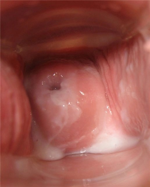
Day eight
The neck is low and closed.
Cervical fluid is white and sticky.
Day nine
The neck is low and closed.
Feeling dry.

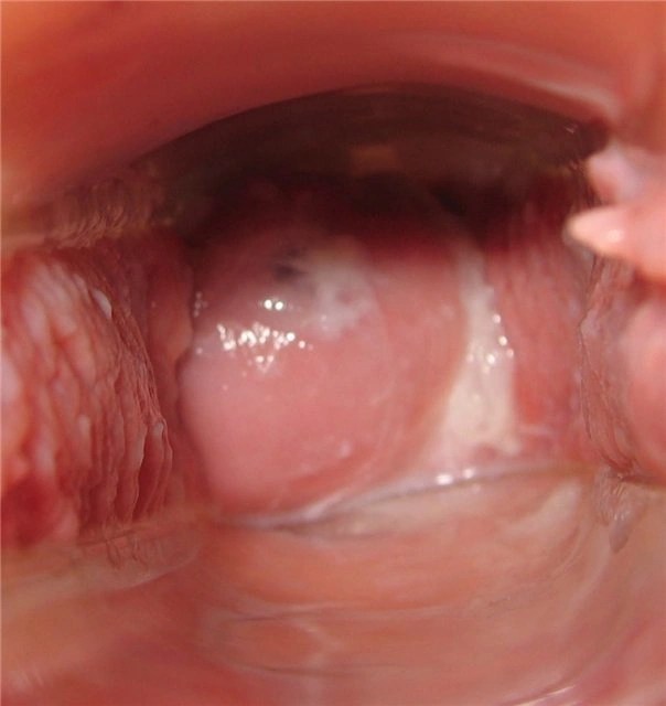
Day ten
The neck is low and closed.
Note the drop of blood and brown lump near the cervix (right). Perhaps from a stormy conversation on the same day, but later an endometrial polyp was diagnosed.

Day eleven
Cervical fluid is creamy.

Day twelve
Cervical fluid is white milky. Feeling wet.
I feel especially sexy.

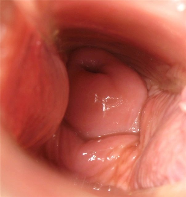
Day thirteen
Copious watery discharge.
The neck is softened and moves upward.

Day fourteen
White, transparent, watery cervical fluid that stains underwear.

Day fifteen
The cervical fluid changes to a discharge that resembles egg white.
The neck is soft, open and high.

Day sixteen
Cervical fluid in the form of egg whites, very wet.
The neck is soft and high.

Day seventeen
Cervical fluid is very thin, with whitish-yellow streaks. Sensual breasts, but not painful.
The liquid stretches between the fingers when stretched.

Day eighteen
Egg white.


Day nineteen
Egg white with a white tint.

Day twentieth
Minor back pain and cramps on the left side.
Suspicion of ovulation.
Feeling of strong sexuality.
Cervical fluid like gelatinous egg white.

Twenty first day
Cervical fluid is like glue.
The nipples are very sensitive and painful.

Day twenty two
Painful nipples.
The neck is in the middle position and slightly open.
Basal body temperature begins to rise.

Day twenty-three
Very sensitive nipples.
Feeling dry.

Day twenty-four
Very sensitive nipples.
Dry.
The neck is firm and high.

Day twenty five
Headache and fatigue.
Cervical fluid is dry/sticky.

Day twenty six
Breasts are swollen.
Basal body temperature is now noticeably higher, by about 1 degree.
Day twenty seven
Painful nipples, swollen breasts.
Cervical fluid is sticky.

Day twenty eight
Feeling dry.

Day twenty nine
Feeling dry.

Day thirty
Feeling dry.
The chest is heavy.

Day thirty one
Feeling bloated.
Dry, (note, fresh blood, a sign of impending menstruation).
Feeling of emotional instability.

Day thirty two
Light brown spots.
The neck is low and open.
Feeling tired.

Day thirty three
Pink spots.
Pain in the lower back.
Menstruation will begin tomorrow after waking up, 13 days after ovulation.
The article was taken from the internet! For those who are not interested, don’t fu... fuck!
Self-diagnosis of pregnancy in the early stages causes certain difficulties. How to determine pregnancy by the cervix, if some girls do not know where it is and what it should look like in its normal state. Reviews and topics on forums speak about this. Even if, if conception is suspected, the woman herself does not intend to detect changes in the main reproductive organ, it is important to know about all her changes, which doctors use as a guide. The most accurate diagnosis will be from a gynecologist.
What is the complexity of the method?
The female body is designed in an amazing way - immediately after fertilization of the egg, the active growth of the fertilized egg begins and moves into the uterus. Active hormonal and physiological changes immediately begin - the woman prepares for the successful bearing and birth of a child. But how can you independently determine pregnancy by looking at the cervix, even before going to the antenatal clinic?When examined by a gynecologist, you can even determine the gestational age by touch - the specialist uses palpation to determine the size of the organ with the embryo growing inside. You can give a more precise date if you keep a cycle chart where the days of ovulation are marked. At home, self-diagnosis will only be approximate. It is necessary to have at least a general idea of the size and shape of the cervix, its density and color before conception and after the fact, as in the figure.
Not all women, even those who have given birth, have a complete understanding of the internal genital organs and how they work. What is the role of each reproductive segment in PA, during fertilization and gestation? If you do not have this basic knowledge, it is difficult to understand how to determine pregnancy by the cervix.
Looking into yourself “there”, even with a mirror, is problematic, especially for overweight ladies. The only way to compare the cervix before and after pregnancy is to feel yourself in the vagina during hygiene procedures to compare the changes.
Attention: This type of diagnosis is very accurate, but it is also considered in terms of sensations and symptoms. Due to the difficulty of conducting a self-examination, it is rarely used even by those who know how to determine pregnancy themselves by looking at the uterus.
Where is the cervix located?
The uterus is an internal organ and is therefore not visible. The lower part of the cervix extends into the vagina; this is the visible part, which is used to make a visual diagnosis of the organ. It is tightly rooted in the vagina, so all sensations are transmitted from the walls of one organ to another (during PA and touching).You can detect pregnancy by palpation by the uterus, and visually by the cervix. The internal cavity of the uterus constantly produces mucus, including spotting during menstruation. A plug is formed in its neck, clogging the internal organ to protect against infections and moisture from the external environment.
Attention: Do not think that the cervix is a secondary organ; the level of protection of the fetus and its retention during pregnancy depends on its condition. If it has lost firmness and elasticity, the doctor, upon examination, can determine an upcoming miscarriage and take measures to preserve the pregnancy.
The specialist also knows how to determine pregnancy with uterine fibroids (internal neoplasm from pathological tissue proliferation). During a visual examination, the doctor can evaluate only the cervical part, but this is enough to assess the health of the entire reproductive organ.
The cervix has the simplest structure - a rounded muscular body, slightly protruding in the upper part of the vagina. It differs in tissue structure and color from the vaginal walls. This pinkish lump is covered with mucus and has a small hole in the center - the cervical canal. It is closed in the normal state, but expands slightly during menstruation.
The passage to the uterus is filled with a mucus plug. The size of the cervix is small - approximately 2.5 cm in circumference to 4 cm in length. It's amazing how this miniature light pink "tunnel" opens and widens during childbirth to allow the baby's head to emerge into the passage!
During ovulation, the mucus plug liquefies so that the most active sperm can overcome this barrier. The cervix rises slightly and becomes softer, making the vagina more free for penetration of the male organ.
How to detect pregnancy by touch
Every gynecologist knows how to determine pregnancy by the uterus, even in the early stages - the lower part of this organ is informative. It shifts, the color, size and density of the tissue changes, they say that the cervix can be soft and “oaky”. These changes are considered the most significant signs of pregnancy, along with the absence of menstruation on time. In addition, traces remain on the cervix:- transferred operations;
- abortions and miscarriages;
- safe birth;
- internal uterine pathologies.
A specialist can easily diagnose the accomplished fact of fertilization, even the approximate gestational age. In nulliparous women, this pharynx is small and rounded; after childbirth, it closes like a slit. After a caesarean section, the cervix is more similar to the cervix of a nulliparous woman, although the cervix becomes slightly larger in size.
You need to know about this before determining pregnancy by touch in the uterus:
- In women, before pregnancy, the cervix is hard, approximately like the wings of the nose; after conception, it is softer, approximately like lips.
- Before pregnancy, the cervix has a velvety pink color, after which it turns blue (from active blood circulation and the proliferation of the vascular network in order to actively supply the fetus with nutrients).
- Under the influence of progesterone (hormone), the cervix lowers - a consequence of completed fertilization.
What changes occur in the cervix after conception?
Only a specialist can determine minor deviations in the condition of the reproductive organs. There are individual characteristics of the body and pathology, but usually you have to focus on average indicators before determining pregnancy by the cervix. It is very difficult to assess tissue density on your own without medical education and palpation experience.Attention: If something “appears” during a self-examination, do not rush to inflate your fantasies and make a diagnosis for yourself! Up to 6 weeks, it is difficult to understand by self-feeling whether you are pregnant or not.
Even if there is a pathology, this should be dealt with by a specialist who can really determine the condition of the reproductive organs. For example, a too hard cervix may indicate hypertonicity (muscle tension) and may “signal” an impending spontaneous miscarriage. This rarely happens in early pregnancy, so don't panic after feeling it. The best way to avoid pregnancy loss is to go to the nearest medical center.
During the examination, the specialist will pay attention to other signs of pregnancy:
- Blueness of the cervix and vaginal walls.
- Slight swelling of the external genitalia.
- Changes in the size, shape and consistency of the uterine walls (rounded and enlarged, becoming soft, called the “Horwitz-Hegar symptom”) over a period of 4–6 weeks.
- After conception, the uterus becomes easily excitable, prone to sudden contractions, becomes dense and sags when examined with both hands - from the vagina and from the abdominal side, this is “Snegirev’s symptom”, a little later it takes its primary position.
- Some mobility of the cervix or “Gubarev-Gaus symptom”, some women have a “Genter symptom”, this is a forward deviation of the uterus with a comb-like thickening in the center.
- Uterine asymmetry or “Piskáček’s sign” is observed in a bicornuate uterus, with one horn slightly larger than the other - a normal phenomenon as long as the embryo develops on one side of the organ. It will become rounder over time, around the 8th week of pregnancy.
Condition of the cervix before childbirth
The cervix changes dramatically before childbirth (and almost imperceptibly for the expectant mother herself) - and this is what allows the child to be born unhindered. What exactly changes does the uterus undergo before childbirth, and in what cases is medical intervention necessary?
What should the cervix look like before natural childbirth?
What matters is its location in the pelvis, length and softness. The fact that a woman’s birth canal is prepared is indicated by the softening of the cervix before childbirth to such an extent that it begins to allow 1-2 fingers of the doctor inside. Due to such changes, the woman observes the release of the mucous plug. It turns out that the earlier the cervix begins to dilate before childbirth, the sooner the woman notices this clear sign of approaching contractions.
In addition, it is shortened. It is known (this is recorded using transvaginal ultrasound) that the length of the cervix before childbirth is no more than 1 cm. And it gradually smoothes out completely.
As for the location, it becomes exactly in the center of the small pelvis, while the cervix during pregnancy, even 3-4 weeks before the onset of labor, is deviated posteriorly.
These 3 parameters are assessed on a two-point scale. With 5 points scored, the cervix is considered mature.
How to stimulate uterine dilatation using medical methods
If an examination of the cervix before childbirth shows that it is not yet mature, whereas, judging by the doctors’ calculations, you should be about to give birth, it is quite possible that you will be asked to speed up the process a little and carry out stimulation. Otherwise, there is a high probability that the child will suffer from oxygen starvation, since by 40-42 weeks the placenta can no longer properly perform its functions; it “grows old.” Stimulation by medical means is possible in a hospital setting. 4 methods are used, sometimes in combination with each other.
1. Intramuscular injections of Sinestrol. This drug speeds up the preparation of the cervix for childbirth, but does not directly provoke contractions.
2. Introduction of kelp sticks - seaweed - into the cervix. This procedure is performed by a doctor, while the patient is on a gynecological chair. The sticks, 5-6 cm each, are placed almost the entire length into the cervical canal. After approximately 3-4 hours, they begin to swell under the influence of moisture, thereby mechanically opening the cervical canal. Within a day, a dilatation of 1 cm is usually observed - a softened and short cervix before childbirth is the key to a quick and easy delivery.
3. Introduction into the cervical canal of a gel containing prostaglandins. For example, Prepidil-gel. It usually acts very quickly, the neck opens in a few hours.
4. Intravenous administration of the drug Enzaprost containing prostaglandins. With its introduction, the cervix becomes soft before childbirth, thereby shortening the period of contractions and expulsion of the fetus.
Self-induction of labor
More often, these techniques are used by women without indications and can be dangerous.
1. Cleansing enema. It is noticed that after it the mucous plug quickly comes away and the cervix opens. This can only be done for those who have already reached their expected due date, that is, the baby is definitely full term.
2. Taking a warm bath. This is not possible if the mucus plug and amniotic fluid have already drained. In addition, it is not recommended for women with high blood pressure.
3. Sex. Sperm contains prostaglandins - the same substances that are part of the drugs used to stimulate labor in a hospital setting. Those who have already lost their mucous plug should not have sex, as there is a high probability of introducing an infection into the uterus. Well, having sex with a condom is useless in terms of stimulating the dilatation of the cervix.
4. Physical activity. Walking up and down stairs, mopping floors while squatting, cleaning the house, etc. But don't overdo it. Especially if you have hypertension or gestosis, or placenta previa.
Now you know what the cervix should be like before giving birth. Just don’t try to independently diagnose how ready she is for childbirth. Leave this task to the doctors.
The cervix changes before menstruation, and it is this feature that makes it possible to determine the imminent onset of menstruation. Some women practice self-examination, but for this you need to know the rules, maintain hygiene, and still visit a gynecologist at least twice a year.
Self-examination
The cervix is a hollow organ that connects the uterus and vagina. You can feel the organ yourself with your fingers by inserting your middle finger into the vagina to its full depth. Tactilely it feels like a bulge.
During menstruation
The cervix becomes loose and soft before the onset of menstruation. The channel is widened, the tip of the finger passes through it. It is wider in those women who have given birth.
For this reason, on the days of menstruation, it is important to especially carefully observe personal hygiene and change the tampon or pad on time. This will prevent infection and pathogenic bacteria from entering.
During pregnancy
What does the cervix look like during pregnancy? It is in the maximum raised position in the vagina, and during palpation it can only be felt with the very tip of the finger.
During pregnancy, the organ becomes dense, hard, and the canal takes on the appearance of a small flat slit.
Is it possible to do self-diagnosis?
Many women, knowing what the cervix should look like before menstruation, when it descends, when it changes its structure, strive to palpate the organ themselves.
And yet gynecologists are against such “examinations”. Let's find out why:
- The presence of a hole in the organ is always a risk of infection entering the uterine cavity. This is especially dangerous if hygiene is not observed. Consequences - inflammatory and infectious diseases, ovarian damage, infertility.
- The position of the organ changes on different days of the menstrual cycle. On some days it is located high, on others it is much lower. Without the necessary information, a woman can penetrate too deeply and damage the organ, which is fraught with infections and the development of erosion.
Still, it is impossible to independently assess the condition of such an important organ, even after gaining experience. It is better if a qualified doctor does this, because when making diagnoses, he relies not only on visual examination and palpation data, but also uses special tools.
In addition, to confirm various assumptions, for example, pregnancy or any diseases, tests will be required.
Video about the structure of the organ
Erosion of the cervix is manifested by dysfunction and destruction of the upper layer of the epithelium, which can only be seen during a gynecological examination
A defect in the cervical epithelium is detected during a routine examination or after a woman contacts a gynecological consultation with complaints of other disorders. The doctor sees changes by examining the patient using mirrors.

Cervix
The vagina and the uterus, hidden inside the woman’s pelvis, are connected to each other by the cervix. From the vagina one can see her external os, which is normally covered with pink epithelium. There is a canal inside the cervix, and the wall of the organ consists of three layers:
- external;
- muscle, consisting of fibers arranged in a circle;
- inner, epithelial layer.
Each of them has its own purpose. Muscular layer - provides compression and sealing of the cervix, protecting the uterus and its contents from infection and fluids.


The inner layer lining the uterine canal consists of a single layer of cells that have a specific, cylindrical shape. They are painted in a specific red color and form a velvety surface; the purpose of such cells is to secrete mucus, which tightly clogs the cervix. The epithelium covering the cervix from the vaginal side has a different structure. Its cells:
- flat, with dense walls, a core inside;
- arranged in several layers;
- tightly closed.


In the lowest, basic row of cells, located at the border of the muscle and epithelial layers, a continuous process of division of existing cells and the birth of new cells occurs. Layers of “fresh” cells rise, squeezing out the aging ones, ensuring complete renewal of the mucous membrane over a short period of time. This process ensures the healing of large and small damage that occurs on the mucosa during life.
The period of such renewal is individual for each woman and depends on the condition of her body.
In some cases, there is a disruption in the functioning of the layer of cells, they cannot be restored as quickly as necessary to maintain their integrity, and ulcerations appear on the cervix. The base layer of epithelium becomes too thin and weak for infection. Such destruction is defined as cervical erosion.
Types of erosion
Patients whose doctor has diagnosed the disease are interested in what cervical erosion will look like (photo), erosion, what is it?
Externally, the damage takes the form of scratches or rounded wounds covering the mucous membrane of the cervix, and defects found on the mucous membranes are usually red.
True erosion
Wounds occur in varying depths and sizes. If lesions:
- observed on the upper epithelial layer;
- do not penetrate deep;
- leave the basal layer intact;
- no infection was detected on the mucous membranes and there was no pronounced inflammation;
- with a fairly strong body, they will be able to grow over themselves, the upper layer of the epithelium will be restored.
When pressing on the wound with an instrument, the doctor may observe slight bleeding - this condition is called true erosion. A doctor can rarely see such a lesion - usually defects of this kind heal within 10-14 days.
If the damage is deep, reaching the basal layer of cells or penetrating below, healing can proceed in two ways:
- a scar will form at the site of the damage, which will make the cervix more rigid and prevent it from stretching during childbirth, which may result in its rupture;
- the defect will not be overgrown with flat epithelium, characteristic of the cervix, but with cylindrical, “velvet” epithelium, which normally should not leave the internal canal of the cervix. This epithelium is located in one layer and produces thick mucus, which is not typical for the cervix.
Pure “erosions” are quite rare. The photo shows erosions of the uterus, which in most cases are accompanied by inflammatory processes or arise against their background.
Causes of the condition
Erosion is a complex condition, the exact mechanism of which is unknown.
Clinical observations made it possible to identify the causes that may provoke a defect in the cervical mucosa. These include:
- damage to the body by sexually transmitted infections that weaken the woman’s body (gonococci, HPV, trichomonas, staphylococci, yeast);
- lowering the body's immune threshold;
- traumatic injury to the cervix due to birth trauma, uterine curettage, surgical treatment or mechanical trauma;
- damage to the epithelium during too early or too late childbirth - during early labor the mucous membrane is still immature, which leads to its destruction;
- inflammatory diseases of the vagina or cervical canal, in which massive mucus secretion and mechanical irritation of the cervical epithelium occur;
- hormonal imbalance due to diseases of the thyroid gland, adrenal function, age-related changes, ovarian tumors;
- violation of the vaginal microflora, which is caused by poor hygiene, unreasonable change of sexual partners;
- discharge from the uterus caused by destruction of fibroids or endometritis, which leads to chemical damage to the cervix and the appearance of wounds.


The resulting condition provokes inflammation of the internal genital organs, weakening the body and can cause infertility.
External signs of the disease
It is believed that erosion has no symptoms and does not manifest itself in any way. But the process is usually accompanied by an infection, which gives cause for concern and consultation with a doctor. The woman begins to be bothered by:


- pain and heaviness in the lower abdomen;
- the appearance of bloody discharge after sexual intercourse;
- pain during sex;
- the appearance of a brownish leucorrhoea (indicates mechanical injury to the cervical mucosa);
- with a long process, mucous leucorrhoea with a purulent odor appears;
- the appearance of a yellowish leucorrhoea indicates the addition of an opportunistic infection;
- Milky white leucorrhoea indicates candidiasis (thrush).
All these conditions, if treated in a timely manner, are treatable and require correction, as they can become a background for malignant changes.
False erosion
False erosion or ectopia is understood as the overgrowing of a defect in the mucosa with single-row columnar epithelium. When you press on the affected area, no blood will be released. The mechanism of replacement of the true epithelium of the cervix with cylindrical epithelium is not completely known. It is believed that the main provoking factor is a hormonal imbalance, which had an impact on the body at the time of formation or in the process of life.


When talking about uterine erosion, most gynecologists mean pseudo-erosion. Women under 30 years of age are most susceptible to this disorder. False erosion can be acquired or congenital.
The doctor can observe in the mirrors what congenital cervical erosion looks like - this is a displacement of the natural pink squamous epithelium, onto which a layer of red velvet cylindrical cells creeps.
Depending on the type of growth, there are several types of pseudo-erosions that differ in appearance.
Glandular
With this lesion, erosion is characterized by greatly overgrown glands - the red epithelium penetrates into the deep layers of the cervical epithelium and “slides” to cover large surfaces. Such a lesion can develop in a woman’s body for a long time (over several years). Clinically, it is the most common type of false erosion.
Papillary


This species is externally designed as symmetrical, clearly shaped growths, reminiscent of miniature papillae, on which a layer of “velvet” epithelium is formed on top. Some doctors attribute this false erosion not to a disease, but to a pathology in the development of tissues of the internal genital organs of a woman.
With the development of such a disorder, a woman may complain of:
- weak nagging pain in the lower abdomen;
- itching and burning in the vagina;
- slight bleeding after sexual intercourse;
- profuse leucorrhoea without a strong odor, white or translucent.
If the leucorrhoea acquires a pungent odor or its color changes, this will indicate an infection.
Cystic false erosion
Such erosion on the cervix (can be seen in the photo) is characterized by the appearance of small cystic growths that form between the layers of the cylindrical epithelium. The cysts are dense and elastic when pressed. This form of change in the integumentary epithelium of the cervix is the most common.
Combined false erosion
The pure, “classic” type of erosion is not so common. In clinical practice, gynecologists most often encounter cervical erosion, which looks like a combination of two types of false erosions - glandular and cystic.
Upon visual inspection, it looks like a combination of enlarged glands and cysts between them. This combination leads to a delay in the secretion secreted by the glands and the addition of infection. The appearance of such an inflammatory process leads to an increase in painful symptoms and will be a reason for a woman to consult a doctor.
Healing false erosion
After correct identification of the underlying disease and its effective treatment, you can observe a process called healing false erosion. The process occurs in a mirror order to the process of the occurrence of false erosion - flat epithelial cells begin to displace the red columnar epithelium, eventually completely replacing it. But if a woman is diagnosed with changes in the epithelium with the appearance of cysts, then they will remain in most cases. Cystic formations enlarge the cervix and change its natural shape.
Papillary form
The erosions on the cervix shown in the photo are papillary growths, covered on top with a layer of columnar epithelium. They contain infiltrated and inflamed areas. The cause of the growths is the human papillomavirus.
Such a disorder can exist for quite a long time on the cervix without significantly disturbing the woman. In this case, dysplasia may develop, which is considered a precancerous condition. Overgrowth of the cervix does not allow sperm to penetrate into the organ cavity, which threatens the development of infertility.
Condition of the cervix after treatment
When carrying out conservative treatment, the condition of the mucous membrane can change - its layer is restored or a scar is formed. After removing the inflammatory processes, to accelerate the restoration of the squamous epithelium layer, cauterization of the affected area is used.
After the procedure, epithelial restoration processes are activated - leukocytes and other cells are drawn into the burn area, enhancing regeneration.
At the site of erosion, a crust forms, which falls off within 10-15 days, revealing part of the mucous membrane, which will fully recover within 45-60 days. When using the radio wave method, not a scab, but a thin film appears on the surface of the treated area.
To accurately diagnose the condition of the cervical cells, it is necessary to examine them under magnification. For this purpose, colposcopy is used, a method that involves examining the organ under a microscope (with the possibility of additional lighting) and a mirror.
To ensure a quality examination, the doctor cleans the cervix. To do this, the vagina is treated with a cotton swab moistened with a solution of vinegar or Lugol.
This treatment makes the lesions more contrasty and allows you to better see the erosion of the uterus and what the lesion looks like.


If, when stained with Lugol, light-colored spots appear on the neck, this raises suspicions of malignant cell degeneration, which requires taking material for examination.
Prevention of the condition
For timely detection of cervical erosion, women over 25 years of age must contact a gynecologist for examination at least once every 6 months for examination by a doctor and a smear.
Important for the prevention of defects in the mucous membrane is the need to choose high-quality underwear that meets the requirements of personal hygiene.
Consistency in choosing a sexual partner will most likely protect you from sexually transmitted diseases and subsequent erosion; it is advisable to practice protected sex even with a regular partner.
In order for the immune system to be strong, you should adhere to a healthy diet, a healthy lifestyle, and treat infectious diseases in a timely manner.